Ilmu Anatomi Tomografi Scan Tomografi Anatomi Tubuh Manusia Scan Tubuh Biology Diagrams
BlogIlmu Anatomi Tomografi Scan Tomografi Anatomi Tubuh Manusia Scan Tubuh Biology Diagrams The data sets were designed to serve as (1) a reference for the study of human anatomy, (2) public-domain data for testing medical imaging algorithms, and (3) a test bed and model for the construction of network-accessible image libraries. The CT data consist of axial CT scans of the entire body taken at 1mm intervals at a pixel resolution Fully annotated brain CT - Normal anatomy of the head on a cross-sectional cranial CT Scan (axial, sagittal and coronal): brain, bones of skull, paranasal sinuses, vasculary territories, cranial base. Menu MY ACCOUNT e-Anatomy Human anatomy atlas IMAIOS DICOM Viewer Free DICOM viewer vet-Anatomy Veterinary anatomy atlas Anatomical structures This article lists a series of labeled imaging anatomy cases by body region and modality. Brain CT head: non-contrast axial CT head: non-contrast coronal CT head: non-contrast sagittal CT head: non-contrast axial with clinical questions CT

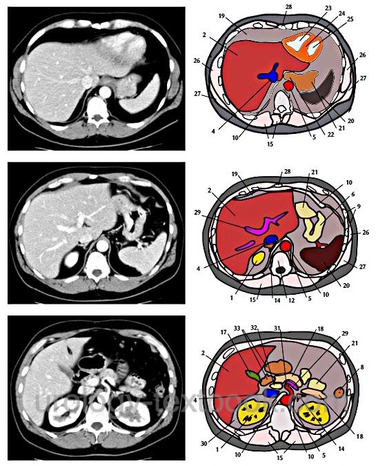
e-Anatomy is a high-quality anatomy and imaging content atlas.It is the most complete reference of human anatomy available on the Web, iPad, iPhone and Android devices. Explore detailed anatomical views and multiple modalities (over 8,900 anatomic structures and more than 870,000 translated medical labels) with images in CT, MRI, radiographs, anatomical diagrams and nuclear images.
Anatomy, the Anatomy of Imaging Biology Diagrams
CT brain - image orientation. Hover on/off image to show/hide findings. Tap on/off image to show/hide findings. Click image to align with top of page. CT brain - image orientation. Roll over the image (tap - mobile devices) to show the annotations; Note the side markers - RIGHT on the viewers LEFT; ANTERIOR brain/head = top of image
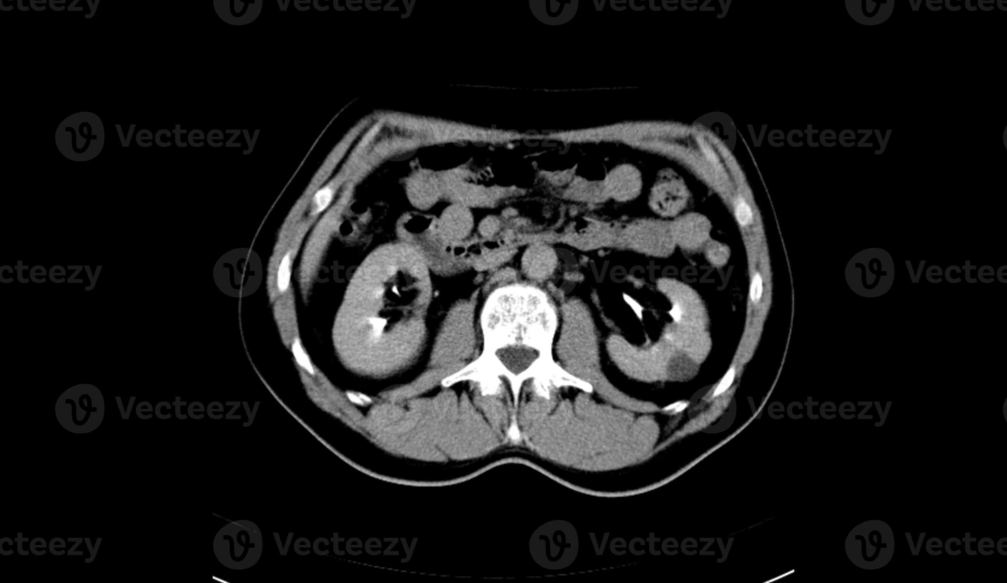
The CT scan brain anatomy is a complex and fascinating field of study, offering a detailed look into the structure and function of the human brain. Through the use of computed tomography (CT) scans, medical professionals can visualize the brain's anatomy, identifying various structures, vessels, and pathways.
Radiology Reference Article ... Biology Diagrams
Normal chest x ray. Radiological anatomy is where your human anatomy knowledge meets clinical practice. It gathers several non-invasive methods for visualizing the inner body structures. The most frequently used imaging modalities are radiography (X-ray), computed tomography (CT) and magnetic resonance imaging (MRI).X-ray and CT require the use of ionizing radiation while MRI uses a magnetic